


Brown to black spots
Brown to black spots observed on tobacco leaves, with examples given below, may have different origins, both biotic and abiotic
 |
 |
 |
 |
 |
| Figure 1 | Figure 2 | Figure 3 | Figure 4 | Figure 5 |
Among the parasitic causes one may note several pathogenic micro-organisms:
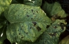
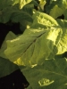
- bacteria such as Pseudomonas cichorii (figure 1), and more uncommonly Pectobacterium carotovorum subsp. carotovorum (Pectobacterium spp.)
- fungi such as Alternaria alternata (figure 2a), Thanatephorus cucumeris (Rhizoctonia solani anam.) (figure 2b), and Corynespora cassiicola, Curvularia verriculosa, Phyllosticta nicotianae and Phytophthora nicotianae. The presence of Alternaria alternata is characterised by the development of velvety black spots formed on conidiophores and conidia of the fungus. Thanatephorus cucumeris is characterised by a greyish white hymenium at the periphery of the spots;
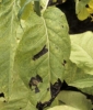
- viruses such as potato virus Y (PVY), tobacco ring spot virus (TRSV) (figure 3), tomato spotted wilt virus (TSWV), tobacco streak virus (TSV), and tobacco necrosis virus (TNV) (see the fact sheet Other parasitic tobacco viruses);
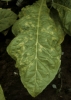
- the nematode Aphelenchoides ritzemabosi (figure 4).
Some non-parasitic diseases may cause brown spots, in particular various nutritional disorders or chemical injuries.
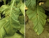
The symptom of partial browning of the midrib and secondary veins is observed when plants are approaching harvest (figure 5). This symptom is frequently but wrongly attributed to the effects of several species of Pectobacterium or PVY. In most cases, it is likely that the saprophytic micro-organisms, namely bacteria, benefit from the leaf maturity to invade the veins. In rainy or foggy periods, after a sprinkler irrigation, water commonly tends to be retained and accumulate on the veins, which facilitates the development of these bacteria.