


Parasitic problems
Pests of air-cured tobacco leaves
Air-curing of tobacco is made without supply of energy in ventilated barns. This curing method lasts much longer than hot air curing (flue-curing) and it of course depends on the curing temperature, and the moisture content in the barn. Note that high humidity sometimes encountered during air-curing can be very favourable for the development of several micro-organisms.
 |
 |
 |
 |
| Figure 1 | Figure 2 | Figure 3 | Figure 4 |
Fungi
Many fungi are likely to cause severe damage in the curing barns:
- Alternaria alternata is rarely observed;
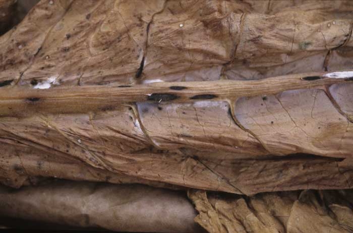
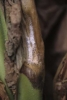
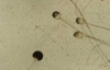
- Botrytis cinerea, a weak parasite can attack tobacco at all stages of development and curing. It causes damage which is often associated with the production of grey mould covering the damaged areas (figure 1);
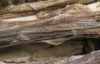
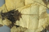
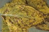
- Botryosporium spp., these saprophyte fungi are easily recognizable by the presence of multitude of long airborne conidiophores on damaged tissues. Two species have been reported on tobacco Botryosporium pulchrum Corda (1840) and Botryosporium longibrachiatum (Oudem.) Mayor (1903). They develop when the tobacco starts turning brown (figures 1 and 2);
- Rhizopus spp. and Mucor spp. are rarely observed on air-cured tobacco. Rhizopus arrhizus var. arrhizus A. Fisch. (1892) is the most frequently reported species. One can also find Rhizopus stolonifer (Ehrenb.) Vuill. (1902) and Rhizopus lyococcus (Ehrenb.) G.Y. Liu, F.L. Lee, Yuan G.F. and Stalpers (2007);
- Sclerotinia sclerotiorum is a dreadful weak parasitic fungus which is frequently observed on air-cured tobacco. Its white mycelium and black sclerotia are characteristic of this fungus presence on damaged tissues;
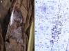
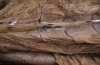
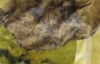
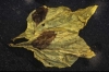
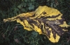
- Cladosporium spp., Fusarium spp., Penicillium spp., and Aspergillus spp., are all saprophytic fungi causing coloured moulds. Note that a number of these fungi may infest the curing barns and warehouses, and in this case, the contamination does not occur in the field (figures 3 and 4);
- Pythium spp., chromista of aquatic environments, especially the species Pythium aphanidermatum.
Bacteria
Some bacteria also affect tobacco leaves in curing barns. Pseudomonas cichorii in France has occasionally been reported. This bacterium only attacks mature tobacco, near or during harvest.
For further information see also this link.
An artificial source of energy is used for leaf drying in case of flue-cured tobacco. This method is used for Virginia type tobaccos. It allows rapid curing of leaves with more precise control of moisture and temperature, the latter ranging from 35 to 70 ° C.
The micro-organisms that sometimes occur during flue-curing are rather different from those observed during air-curing of tobacco.
 |
 |
 |
 |
 |
 |
| Figure 1 | Figure 2 | Figure 3 | Figure 4 | Figure 5 | Figure 6 |
Fungi
Similar to the "air-cured" leaves, several fungi may be responsible for damage caused to flue-cured tobacco leaves.
- Rhizopus spp. and Mucor spp. are frequently observed micro-organisms in curing barns. Rhizopus arrhizus var. arrhizus A. Fisch. (1892) is the most frequently reported species (figures 1 to 4). This saprophytic fungus grows well at high temperatures between 25 and 40 ° C. Note that it tolerates temperatures around 80 ° C for 72 h. Moreover, it has also been reported attacking the stem cortex under field conditions in Nicotiana glauca;
- Alternaria alternata is frequently observed. This weak parasitic fungus occurs also in the field, it can easily develop on mature tobacco leaves (figure 5);
- Botrytis cinerea is more rarely met in flue-curing barns;
- Sclerotinia sclerotiorum occurs only occasionally;
- Cercospora nicotianae and Gibberella tricincta El-Gholl, McRitchie, Schoult. and Ridings (1978), former Fusarium tricinctum (Corda) Sacc. (1886) may cause some damage.
Bacteria
Some bacteria are reported to cause leaf damage in curing barns:
- Pseudomonas syringae pv. angulata has been occasionally described;
-Pectobacterium spp. such as Pectobacterium carotovorum subsp. carotovorum and Pectobacterium chrysanthemi can cause rot spreading from veins (figure 6).
For additional information, please follow this link.