


Internal stem lesions
Some tobacco diseases cause symptoms specifically inside the stem. They can often cause other symptoms in different organs of a tobacco plant, and that it is why it is important to consult the other related topics.
The visible lesions on the stem are essentially caused by various pathogenic micro-organisms:
- bacteria such as Pectobacterium spp. (see also Lesions from wounds) Ralstonia solanacearum;
- and fungi, especially Fusarium oxysporum f. sp. nicotianae, Peronospora hyoscyami f.sp. tabacina or Verticillium dahliae;
- but also various viruses (see for example, tomato spotted wilt virus).
Some rare non-parasitic diseases can damage the internal tissues of the stem, such as injuries caused by lightning (see Lightning injuries).
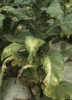
Note that among the micro-organisms previously reported, a number of them are classified as vascular diseases (Fusarium oxysporum f. sp. nicotianae, Verticillium dahliae, Ralstonia solanacearum), which means that they are located and they progress in the plants vessels. The location and vascular proliferation of these pathogens, along with the plant responses (many gummy substances secreted to block the pathogen), also cause other very characteristics, often sectorial symptoms, such as wilting, yellowing and drying of leaves, (figure 1).
 |
 |
 |
 |
| Figure 1 | Figure 2 | Figure 3 | Figure 4 |
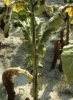
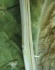
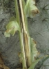
Generally, the colonised vessels turn yellow or brown (figures 2-4); adjacent tissues, such as the cortex or the pith may also be affected. From that moment we start observing the following symptoms outside the stem:
- unilateral longitudinal yellowing of the stem;
- unilateral browning or longitudinal necrotic alteration of the stem;
- browning of a significant portion of the stem.
To identify these vascular diseases one should observe the plants very carefully for the above described symptoms. One will see that these diseases cause often rather similar symptoms on tobacco. In many cases it would be preferable to confirm the diagnosis by a specialised laboratory which could analyse isolation from the vascular tissues using an artificial culture medium.